A Brief History of Sickle Cell Disease. When anemia comes on slowly the symptoms are often vague such as tiredness weakness shortness of breath headaches and a reduced ability to exercise.

Pin On ℂlinic ℒaboratory
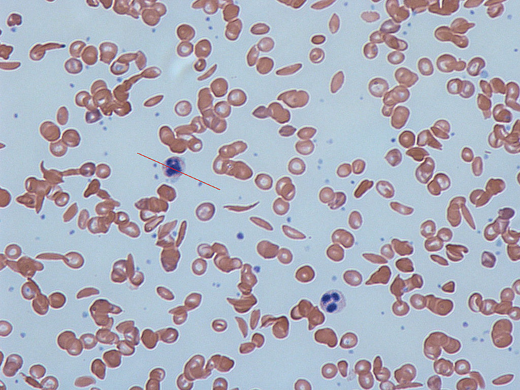
To confirm the diagnosis a sample of blood is examined under a microscope to check for large numbers of sickled red blood cells - the hallmark trait of the disease.

. The blood is examined under a microscope and counted. This is a clinical laboratory finding. Sickle cell disease.
The red blood cells will take on a lighter color when viewed under the microscope. When Herrick saw this in the chart he became interested because he saw that this might be a new unknown disease. To do the count you first have to draw blood from your arm into a clean tube.
In more than 40 states testing for the defective sickle cell gene is routinely performed on newborns. In individuals of the AS genotype such blood cells sickle and are then eliminated by macrophage cells of the bodys immune system lessening the burden of infection Luzzatto 2012. Blood smears can help detect abnormalities in the blood cells such as.
Carriers of the sickle cell. A child is born with the condition of normocytic anemia. Herrick described an anemia characterized by bizarre.
Normocytic anemia can be congenital in some cases ie. Sickle-shaped RBCs may be. In a peripheral blood smear the blood sample is viewed under the microscope.
One example is sickle cell anemia. Sickle cell anemia. With a test called a hematocrit you can.
Neutrophil hypersegmentation can be defined as the presence of neutrophils whose nuclei have six or more lobes or the presence of more than 3 of neutrophils with at least five nuclear lobes. Individuals with anemia and a. Women who have sickle cell disease have red blood cells that change shape and break down more quickly than normal red blood cells.
In addition to being able to visualize differences in size and shape other findings may include target cells nucleated red blood cells fragmented red. Other tests such as examining a blood sample under a microscope and less often examining a sample taken from the bone marrow help determine the cause of the anemia. The decision to begin transfusion depends on the rate of fall of the hemoglobin and the patients clinical condition.
Sickle cell disease can also be detected in an unborn baby. Low levels of hemoglobin or a low hematocrit the percentage of red blood cells in the total blood volume found in a blood sample confirm the anemia. The sickle cell mutation is relevant to malaria because infection of a red blood cell with the malaria parasite leads to hypoxia.
It is visualized by drawing blood from a patient and viewing the blood smeared on a slide under a microscopeNormal neutrophils are uniform in size with an. Viewing the peripheral blood smear under a microscope. These can include blood smears where a film of your blood is examined under a microscope.
While diagnosing for the case of normochromic anemia the normochromic red blood cells appear to be normal in shape and size under the microscope with no characteristic difference as in the case of sickle cell anemia. High RDW and Low MCV. Blood transfusion is necessary for aplastic crisis indicated by low reticulocyte counts.
Sickle cell anemia is an inherited disorder that leads to the production of an abnormal type of. The number and type of red blood cells are evaluated to see if they are normal. Irons examined Noels blood under the microscope and saw red blood cells he described as having the shape of a sickle.
Helmet cells schistocytes RBC fragments and spherocytes. Iron deficiency anemia sickle cell beta-thalassemia or hemoglobin H. Stained layer of blood on a slide under a microscope.
Anemia or anaemia British English is a blood disorder in which the blood has a reduced ability to carry oxygen due to a lower than normal number of red blood cells or a reduction in the amount of hemoglobin.

Pin On School

Pin On Microscopy

Pin On Microscopic Stuff
Pin Em Blood Morphology

Pappenheimer Bodies And Basophilic Stippling In Sickle Cell Disease 3 Sickle Cell Disease Hematology Sickle Cell

Pin On Biology

Pin On Sickle Cell
Pin Em Hematology
0 comments
Post a Comment